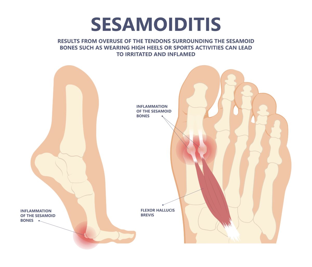
If you’ve ever felt a nagging ache under the ball of your foot, especially beneath the big toe, you might be dealing with sesamoiditis. This condition involves inflammation of the sesamoid bones—two tiny, pea-sized structures embedded in the tendons beneath the first metatarsophalangeal (MTP) joint. Despite their small size, these bones play a crucial role in how you walk, run, and push off the ground. When inflamed, sesamoiditis can turn everyday movements into painful struggles, significantly impairing mobility and quality of life.
Sesamoiditis is typically caused by repetitive stress or overuse, making it common among athletes, dancers, and people who spend long hours on their feet. Unlike an acute injury, it develops gradually, often mimicking other foot conditions such as turf toe or gout. Because the sesamoid bones have limited blood supply, healing can be slow, and without proper care, the condition may become chronic. Understanding the anatomy, symptoms, and treatment options is key to regaining pain-free movement and preventing long-term complications.
This guide explores everything you need to know about sesamoiditis—from the function of the sesamoid bones and common causes of inflammation to how it affects mobility and the most effective ways to treat and prevent it.
How Sesamoid Bones Support Foot Function

Mechanical Role in Gait and Movement
The sesamoid bones act like biological pulleys within the flexor hallucis brevis tendon, enhancing the mechanical efficiency of toe movement. During the push-off phase of walking or running, they increase the force generated by the tendon, allowing for a more powerful and controlled lift of the big toe. This mechanism reduces the workload on surrounding muscles and tendons, making movement smoother and more efficient.
These bones also absorb and distribute significant pressure—up to three times your body weight during high-impact activities like sprinting or jumping. By spreading this load across the forefoot, they protect the joint and prevent excessive strain on soft tissues.
Anatomical Position and Structure
Located beneath the head of the first metatarsal, the two sesamoid bones are known as the medial (tibial) and lateral (fibular) sesamoids. The medial sesamoid is larger and bears more weight, making it more prone to injury. Connected by the intersesamoidal ligament and supported by the plantar plate, they help stabilise the MTP joint during motion.
Unlike most bones, sesamoids are not linked by joints. Instead, they float within tendons—similar to the kneecap—making them uniquely vulnerable to compression and shear forces, especially during activities that involve repeated toe-off or squatting.
Poor Blood Supply and Healing Challenges
A major reason sesamoid injuries heal slowly is their limited vascular supply, particularly in the central and proximal regions. Reduced blood flow means fewer nutrients and oxygen reach the bone, increasing the risk of delayed healing, stress reactions, and even avascular necrosis (bone death). This explains why conservative treatment often takes weeks or months and underscores the importance of early intervention.
Common Causes of Sesamoiditis
Repetitive Forefoot Loading
Activities that place continuous pressure on the ball of the foot are primary triggers. Runners, dancers (especially ballet), basketball players, and gymnasts frequently develop sesamoiditis due to repeated toe-off motions. Baseball catchers and weightlifters who squat deeply are also at high risk because of sustained pressure on the MTP joint.
Even non-athletes can develop the condition from prolonged standing, walking on hard surfaces, or suddenly increasing physical activity without proper conditioning.
Foot Structure and Gait Issues
Certain foot types increase susceptibility:
– High arches (pes cavus) concentrate pressure on the forefoot.
– Flat feet (pes planus) alter weight distribution, overloading the medial sesamoid.
– Excessive pronation causes the foot to roll inward, straining the joint.
Abnormal walking patterns, such as limping or toe-walking, can also create uneven stress, accelerating wear and inflammation.
Inappropriate Footwear
High heels shift up to 75% of body weight forward onto the forefoot. Thin-soled shoes, tight toe boxes, and minimalist footwear offer little cushioning, increasing direct pressure on the sesamoids. Dancers in soft slippers or athletes in cleats may experience similar effects.
Underlying Medical Conditions
- Bipartite sesamoids, a congenital split in the bone, are more prone to irritation and often mistaken for fractures.
- Gout can cause crystal-induced inflammation in the MTP joint, mimicking sesamoiditis.
- Hallux rigidus or bunions change joint mechanics, increasing sesamoid strain.
Recognizing Early Warning Signs

Gradual Onset of Pain
Pain usually starts mild and worsens with activity. It’s often described as a dull ache or throbbing beneath the big toe joint, especially on the inner (medial) side. Early symptoms include discomfort after running or standing—improving with rest but returning with use.
Ignoring early signs can lead to chronic inflammation and tissue damage.
Tenderness and Swelling
Direct pressure on the sesamoid area causes sharp tenderness. Swelling may be subtle, but inflammation is often detectable during clinical exams. Some patients report a “walking on a pebble” sensation due to the inflamed bone pressing against the sole.
Limited Big Toe Movement
As inflammation increases, bending or straightening the big toe becomes painful. This restricts the push-off phase of gait, forcing you to alter your stride. Stiffness may be worse in the morning or after prolonged sitting.
Common Pain Triggers
- Walking barefoot on hard floors
- Climbing stairs or squatting
- Wearing high heels or unsupportive shoes
- Driving (pressure on pedals)
Persistent pain at rest suggests advanced inflammation or a possible stress fracture.
How Doctors Diagnose Sesamoiditis

Clinical Evaluation and Physical Tests
Diagnosis begins with a detailed history and physical exam. Your doctor will assess activity levels, footwear, and symptom progression. Palpation over the sesamoids helps localise pain. The Passive Axial Compression (PAC) test—pressing the big toe along its length while stabilising the metatarsal—reproduces sesamoid pain and helps differentiate it from other conditions.
Imaging for Accurate Diagnosis
X-rays are typically first, ruling out fractures and identifying bipartite sesamoids. Smooth edges suggest a normal variant; jagged lines may indicate a fracture.
However, X-rays can miss early stress injuries. MRI is the gold standard, detecting bone marrow oedema, soft tissue inflammation, and early osteonecrosis. Bone scans show areas of increased metabolic activity, useful for stress fractures. Ultrasound can guide injections, while CT scans help with surgical planning.
Ruling Out Other Conditions
Sesamoiditis must be distinguished from:
– Sesamoid fracture (sudden pain, trauma history)
– Turf toe (ligament injury from hyperextension)
– Bursitis or tendinosis
– Gout (acute, red, hot joint)
Misdiagnosis is common, so a comprehensive evaluation is essential.
Mobility Limitations Caused by Sesamoiditis
Altered Gait and Walking Pain
The first MTP joint is vital during propulsion. When impaired, you may shorten your stride, walk on your heels, or limp. This reduces walking efficiency, increases fatigue, and may lead to secondary issues like plantar fasciitis or Achilles tendinitis.
Reduced Athletic Performance
Runners lose explosive power. Dancers struggle with jumps and relevé. Athletes in sports like soccer or tennis find cutting painful. Studies show only 45.4% return to pain-free sports after treatment.
The Foot and Ankle Ability Measure (FAAM) reveals significant deficits, even after symptom improvement.
Daily Activity Challenges
Simple tasks become difficult:
– Climbing stairs
– Standing in line
– Driving
– Wearing regular shoes
Chronic pain may force lifestyle changes, such as quitting dance or avoiding walks.
Risk of Compensatory Injuries
To protect the injured foot, you may shift weight to the outer edge or other toes, causing metatarsalgia, stress fractures, or ankle instability. Overuse of the opposite limb may lead to knee or hip pain.
Conservative Treatment Options
Immediate Pain Relief Strategies
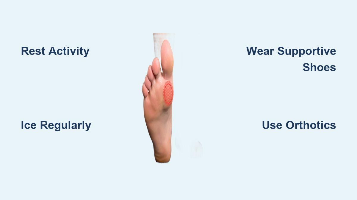
Start with RICE—Rest, Ice, Compression, Elevation:
– Rest: Avoid aggravating activities.
– Ice: Apply 15–20 minutes every few hours.
– NSAIDs: Ibuprofen or naproxen to reduce pain and swelling.
Topical anti-inflammatories are an alternative.
Offloading the Sesamoid Area
Reducing pressure is key:
– Sesamoid cutout orthotics
– Metatarsal pads
– Stiff-soled or rocker-bottom shoes
– CAM boots for moderate to severe cases
Custom orthotics are more effective, especially for high arches or flat feet.
Medical Injections and Therapies
When pain persists:
– Corticosteroid injections (ultrasound-guided) reduce inflammation.
– Platelet-rich plasma (PRP) may promote healing in chronic cases.
– Concentrated bone marrow aspirate (CBMA) for refractory cases.
Space injections at least three weeks apart.
Physical Therapy and Rehabilitation
Restoring Range of Motion
Gentle stretching prevents stiffness:
– Big toe dorsiflexion stretches
– Towel-assisted mobilisations
– Joint mobilisation by a therapist
Avoid aggressive manipulation.
Strengthening Intrinsic Foot Muscles
Weak foot muscles increase sesamoid load. Exercises include:
– Towel scrunches
– Marble pickups
– Short foot exercises
– Heel raises
Advanced Recovery Modalities
- Extracorporeal shockwave therapy (ESWT)
- Laser therapy
- Iontophoresis
- TENS units
Balance and proprioception training help restore natural gait.
When Surgery Is Necessary
Indications for Surgery
Surgery is considered if:
– No improvement after 4–6 months
– Non-healing or displaced fracture
– Severe avascular necrosis
– Pain affects quality of life
Imaging must confirm structural damage.
Sesamoidectomy Procedure
Sesamoidectomy—removal of the affected sesamoid (usually medial)—is the most common surgery. Performed through a small sole incision, it preserves surrounding tendons.
Postoperative Recovery Timeline
- Weeks 1–4: Non-weight-bearing in a cast or boot.
- Weeks 5–12: Gradual weight-bearing, physical therapy.
- Months 3–6: Return to normal shoes, low-impact activities.
- 6+ months: Possible return to sports.
Full recovery can take up to a year.
Long-Term Outlook and Prevention
Healing Time and Prognosis
Most improve in 6–12 weeks, but:
– Mild: 4–8 weeks
– Moderate to severe: 3–6 months
– Chronic cases: up to 22 months
A review found 66% reduced pain, but only 45.4% returned to pain-free sports.
Preventing Future Episodes
- Wear supportive, low-heeled shoes
- Use custom orthotics if needed
- Follow the 10% rule when increasing activity
- Strengthen feet regularly
- Maintain a healthy weight
Even a 10-pound weight loss reduces forefoot pressure.
Final Note: Sesamoiditis is more than foot pain—it’s a mobility-limiting condition that disrupts how you move, work, and play. By understanding its causes, recognising early symptoms, and following structured treatment and prevention strategies, you can protect your feet and maintain long-term function. Whether you’re an athlete, dancer, or simply on your feet all day, proactive care is the key to staying pain-free.