If you’ve ever experienced sharp heel pain with your first steps in the morning, or wondered how your foot can be both flexible and firm during walking, the answer lies in a brilliant biomechanical system known as the windlass mechanism of the foot. This dynamic process transforms your foot from a shock-absorbing platform into a rigid lever at the precise moment you push off the ground, enabling efficient and powerful movement. Without it, every step would require more muscular effort, reduce propulsion, and increase stress on joints and soft tissues.
At the heart of this mechanism is the plantar fascia, a strong, fibrous band that runs along the sole of your foot from the heel to the base of your toes. As you rise onto your toes during walking, your big toe extends upward—dorsiflexes—causing the plantar fascia to tighten like a cable being wound around a drum. This action lifts the arch of your foot and locks the midfoot joints, creating a stable structure ideal for transmitting force. First described by Leroy Root in 1977, the mechanism gets its name from the nautical windlass, a device used to pull in anchor chains, due to its similar winding action.
Understanding the windlass mechanism isn’t just for biomechanics experts—it’s essential for diagnosing and managing common foot conditions like plantar fasciitis, hallux limitus, and flat feet. In this guide, you’ll learn how the windlass mechanism functions, what happens when it fails, how clinicians test it, and what you can do to keep it working properly.
How the Windlass Mechanism Turns Your Foot Into a Rigid Lever
The windlass mechanism is most active during the final phase of walking, turning your foot into a stable, energy-efficient structure for push-off. It’s not passive—it’s a precisely timed, dynamic process driven by movement and anatomy.
Activation During Push-Off: The Moment Your Arch Rises
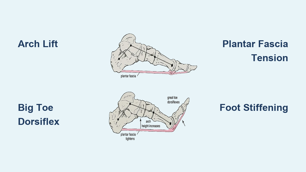
The windlass mechanism engages during terminal stance, just before your foot leaves the ground. As your heel lifts, your body weight shifts forward onto the ball of your foot. The ground resists your toes—especially your big toe—forcing them to dorsiflex (bend upward). This motion pulls the plantar fascia taut over the metatarsal heads, the rounded bones at the base of your toes.
Imagine the plantar fascia as a rubber band stretched between your heel and toes. When you lift your heel and bend your big toe up, the band tightens. This tension shortens the distance between your heel and forefoot, which elevates the medial longitudinal arch and stabilizes the midfoot. The foot transitions from flexible to rigid—exactly what’s needed to push off efficiently.
Without this transformation, your foot would remain too loose during propulsion, making walking less efficient and placing greater strain on muscles and ligaments.
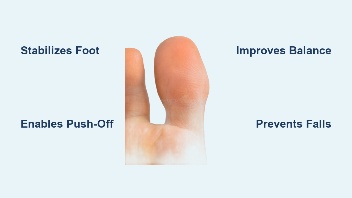
Why the Big Toe Is the Key to Windlass Activation
The hallux (big toe) is the primary driver of the windlass mechanism. It undergoes the greatest range of dorsiflexion—about 54° at push-off, compared to 32–49° in the smaller toes. Positioned under the largest metatarsal head and connected to the strongest portion of the plantar fascia, the big toe generates the most effective arch lift.
If the big toe can’t extend properly—due to arthritis, stiffness, or injury—the windlass mechanism fails to activate fully. This leads to reduced propulsion, compensatory gait patterns, and increased risk of pain and overuse injuries.
Key Structures That Enable the Windlass Mechanism

The windlass mechanism relies on the precise interaction of several anatomical components. Damage or dysfunction in any one can impair the entire system.
Plantar Fascia: The Tension Cable That Lifts the Arch
The plantar fascia (or plantar aponeurosis) originates at the medial tubercle of the calcaneus and fans out to attach beneath the toes. It acts like a bowstring, resisting arch collapse under body weight.
During windlass activation, the fascia experiences tension up to 1.5 times your body weight. The medial slip, which runs beneath the big toe, bears up to 27% of the total load, making it the most stressed portion. This explains why plantar fasciitis often causes pain near the heel attachment of this slip.
Metatarsal Heads: The Pivot Point That Wraps the Fascia
The metatarsal heads serve as the fulcrum—the “drum” around which the plantar fascia wraps. Their curvature influences how much tension is generated. The first metatarsal head has the largest radius (about 9.2 mm), allowing greater leverage and more effective winding of the fascia.
When the toes dorsiflex, the fascia rides up the anterior slope of these heads, increasing tension and lifting the arch. A higher or more prominent metatarsal head enhances this effect, while a flattened one reduces it.
Medial Longitudinal Arch: The Structure That Gets Stabilized
The medial longitudinal arch (MLA) is the highest and most mobile arch of the foot. It’s composed of the calcaneus, talus, navicular, cuneiforms, and metatarsals. Under load, it tends to flatten—unless counteracted by the plantar fascia.
In the windlass mechanism, the fascia acts as a tie-rod, preventing excessive lowering of the arch. This stabilization is crucial for maintaining foot rigidity during propulsion and protecting joints from strain.
The Triangular Truss Model: How the Foot Resists Deformation

The foot functions like an engineered bridge, thanks to the triangular truss model.
How Geometry Creates Stability
In this model:
– The base is the plantar fascia.
– The two sides are the calcaneus and metatarsals.
– The apex is at the metatarsophalangeal joints (MTPJs).
This triangular configuration creates a mechanically stable structure that resists deformation under load. Like a bridge truss, it distributes forces efficiently across multiple components.
When the windlass activates, the base (plantar fascia) shortens, raising the apex and tightening the entire structure. This converts the foot into a rigid lever, ideal for force transmission during push-off.
Windlass Mechanism in the Gait Cycle: Timing Is Everything
The windlass mechanism is precisely timed within the gait cycle to support each phase of walking.
Phases of Gait and Windlass Activation
- Heel Strike: Foot lands slightly supinated. Plantar fascia is slack.
- Mid-Stance: Foot pronates to absorb shock. Arch lowers slightly.
- Terminal Stance (Heel-Off): Heel lifts, toes contact ground. Ground forces begin to dorsiflex the hallux.
- Pre-Swing (Push-Off): Hallux extends further, winding the fascia. Arch rises, foot stiffens.
- Swing Phase: Mechanism resets.
The windlass effect begins almost immediately after heel lift. In healthy individuals, arch elevation starts within 0–15° of hallux dorsiflexion. Delays beyond this indicate dysfunction.
Preloading: The Plantar Fascia Is Primed Before Activation
Recent research shows the plantar fascia is preloaded even before heel strike. Muscles like the tibialis anterior and extensor digitorum longus activate early, creating initial tension.
This pre-stretch:
– Removes collagen fiber crimp, enabling faster stiffening.
– Enhances energy storage and return.
– Improves propulsion efficiency.
Studies confirm plantar fascia tension is significantly above rest during early stance (P < 0.01), proving the system is primed before full activation.
Quantitative Biomechanics: The Numbers Behind the Windlass

Understanding the forces involved helps explain why dysfunction leads to pain.
Tension and Strain in the Plantar Fascia
- Maximum tension: Up to 1.5 body weights (BW) during push-off.
- Strain (elongation): Ranges from 3.5% to 6% of resting length.
- Peak strain timing: Occurs at ~80% of stance phase.
The medial slip experiences the highest strain due to greater elongation and length (~156 mm). Lateral slips are shorter (~136 mm) and stiffer.
Metatarsophalangeal Joint Motion
| Toe | Dorsiflexion at Heel-Strike | Dorsiflexion at Push-Off |
|---|---|---|
| 1st | 29 ± 8° | 54 ± 3° |
| 2nd | 29 ± 12° | 49 ± 5° |
| 3rd | 29 ± 8° | 44 ± 2° |
| 4th | 31 ± 10° | 42 ± 2° |
| 5th | 27 ± 10° | 32 ± 7° |
Motion decreases from medial to lateral, reflecting medial-to-lateral sequencing of push-off.
Force Transfer and Joint Stability
A strong linear relationship exists:
Contact Force = 1.13 × Fascia Tension – 0.1 (R² = 0.923)
This means the windlass mechanism stabilizes MTPJs by applying a plantarflexion moment, counteracting upward ground forces. Without it, pressure shifts to metatarsal heads, increasing risk of metatarsalgia.
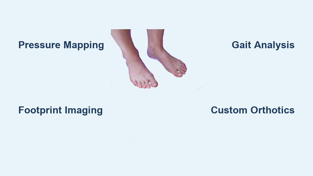
How to Test the Windlass Mechanism Clinically
Clinicians use the windlass test to assess plantar fascia integrity, especially in suspected plantar fasciitis.
Performing the Windlass Test
Weight-Bearing Windlass Test
- Patient stands barefoot.
- Examiner dorsiflexes the hallux.
- A positive test reproduces heel pain.
This version is more physiologically relevant.
Non-Weight-Bearing Test
- Patient sits.
- Examiner passively dorsiflexes toes.
- Less sensitive, but useful for screening.
Diagnostic Accuracy
| Test Type | Sensitivity | Specificity |
|---|---|---|
| Weight-Bearing | Higher | 100% |
| Non-Weight-Bearing | 32% | Not reported |
De Garceau et al. (2003) found 100% specificity—a positive test almost certainly indicates plantar fascia dysfunction.
Final Note: The windlass mechanism is a masterpiece of biomechanical engineering, enabling efficient, stable, and pain-free movement. When it fails, the consequences are felt in every step. By understanding how it works—and how to support it—you can prevent and treat common foot problems, maintain mobility, and keep walking strong.