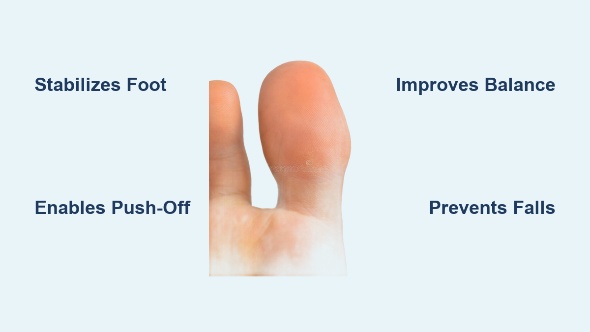
If you’ve ever experienced foot pain after a long day on your feet—or wondered why certain shoes feel better than others—the answer may lie in how your foot interacts with the ground. A podoscope is a cutting-edge diagnostic device that reveals exactly that: it captures detailed images and measures pressure distribution across the sole of the foot, offering clinicians an objective view of foot function. More than just a footprint scanner, modern podoscopes combine high-resolution optical imaging with real-time pressure sensor technology to analyze both the structure and biomechanics of the foot under weight-bearing conditions.
Used in podiatry clinics, sports medicine labs, diabetes care centers, and orthopedic practices, the podoscope transforms subjective observations into quantifiable data. It helps detect flat feet, high arches, gait imbalances, and early signs of conditions like plantar fasciitis or diabetic ulcers—often before symptoms worsen. In this guide, we’ll explore what a podoscope is, how it works, and exactly how it measures foot pressure to support faster diagnoses, personalized treatments, and preventive care.
How a Podoscope Captures Foot Structure and Function
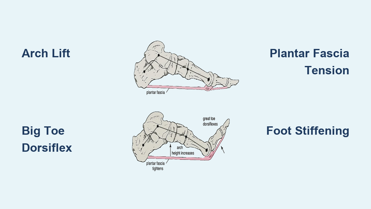
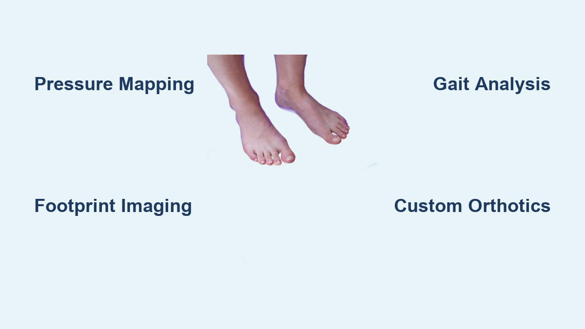
At its core, a podoscope is designed to visualize the plantar surface (the bottom) of the foot while it’s bearing weight. Unlike X-rays or MRI scans, which show internal structures, a podoscope focuses on how the foot actually performs when standing or walking. This functional insight is essential because many foot problems only become apparent under load.
The device typically consists of a translucent platform illuminated from below, equipped with high-definition cameras and pressure-sensitive sensors. When a patient stands barefoot on the surface, light reflects differently depending on where the foot makes contact. Areas in full contact absorb more light and appear darker; areas that don’t touch the platform reflect more light and show up as brighter zones. This contrast creates a sharp, high-contrast image of the footprint, revealing arch shape, weight distribution, and alignment.
Static vs. Dynamic Scanning: Two Views of Foot Health
Modern podoscopes offer two types of analysis:
- Static scanning records foot posture while the patient stands still. It shows:
- Arch type (normal, flat, or high)
- Symmetry between left and right feet
-
Overall contact area and pressure zones
-
Dynamic scanning tracks changes during walking or movement. Found in advanced systems like Podoflash 2.0 and Footwork Lab, dynamic mode captures:
- Center of pressure (COP) trajectory
- Timing of heel strike and toe-off
- Abnormal pronation or supination patterns
Dynamic analysis is especially valuable for athletes, diabetic patients, and those recovering from injury, as it reveals gait mechanics that static images alone cannot detect.
The Science Behind Foot Pressure Measurement
While visual imaging shows foot shape, pressure sensors reveal how force is distributed across the sole. This is where the podoscope becomes a biomechanical powerhouse.
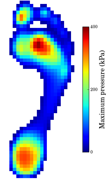
Embedded within the scanning platform are hundreds of capacitive or resistive sensors, each capable of detecting minute changes in force. As the foot presses down, these sensors generate electrical signals proportional to the amount of pressure applied. The data is then translated into a color-coded pressure map, similar to a weather radar, where:
- Red and orange indicate high-pressure areas (e.g., under the heel or ball of the foot)
- Yellow and green represent moderate pressure
- Blue and purple show low or no contact
These thermal-style maps allow clinicians to instantly identify “hot spots” where excessive pressure could lead to pain, calluses, or even tissue breakdown—critical for preventing diabetic foot ulcers.
Types of Pressure Sensors Used
Different podoscopes use various sensor technologies:
- Capacitive sensors: Measure changes in electrical capacitance when compressed. Highly accurate and durable, ideal for clinical use.
- Resistive sensors: Detect shifts in electrical resistance under pressure. Common in entry-level systems due to lower cost.
- Piezoelectric sensors: Generate voltage in response to mechanical stress. Best suited for dynamic gait analysis due to fast response times.
Each sensor corresponds to a pixel in the final pressure map, enabling millimeter-level precision in identifying problem zones.
Analyzing Gait and Load Distribution Over Time
Advanced podoscopes don’t just take a snapshot—they record how pressure shifts during movement. This dynamic load analysis is key to diagnosing gait disorders and optimizing athletic performance.
Tracking the Center of Pressure (COP)
The center of pressure (COP) is the point at which the total force from the foot is concentrated. As a person walks, the COP should follow a smooth, predictable path from heel to toe.
Abnormalities in COP movement can signal:
– Overpronation: Excessive inward rolling, often linked to flat feet and knee pain
– Supination: Outward rolling, associated with ankle instability and stress fractures
– Irregular trajectories: May indicate neurological issues or pain-avoidance behaviors
Software tools plot COP path length, speed, and variability, giving clinicians measurable benchmarks for tracking treatment progress.
Time-Based Pressure Graphs and Gait Phases
Dynamic systems display pressure over time using waveform graphs or pressure bars:
– X-axis: Time (from heel strike to toe-off)
– Y-axis: Force (measured in kPa)
These graphs reveal:
– Delayed or early push-off phases
– Asymmetrical loading between feet
– Instability during midstance
For runners or dancers, this data pinpoints inefficiencies in stride timing and balance, helping prevent overuse injuries.
Correlating Data with Shoe Wear Patterns
Some systems allow integration with abrasion maps—digital scans of worn shoe soles. By comparing external wear patterns with internal pressure data, clinicians gain deeper diagnostic confidence.
For example:
– Heavy wear on the outer heel → confirms supination
– Forefoot wear under the big toe → suggests hallux limitus or bunions
This dual-source analysis enhances accuracy in orthotic design and footwear recommendations.
Key Diagnostic Applications in Clinical Practice

Podoscopes are used across specialties to diagnose, treat, and prevent foot-related conditions.
Early Detection of Diabetic Foot Ulcers
For diabetic patients with neuropathy, foot ulcers often develop silently. The podoscope identifies high-pressure zones before skin breakdown occurs, enabling preventive offloading with custom insoles. Studies show pressure-guided interventions can reduce ulcer recurrence by up to 50%.
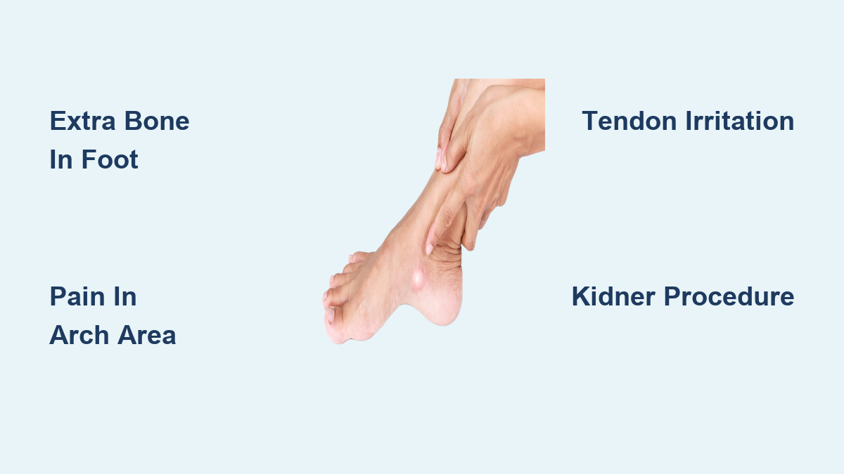
Managing Plantar Fasciitis and Heel Pain
Patients with plantar fasciitis typically exhibit elevated pressure under the medial heel and restricted ankle motion. Podoscopic data guides targeted treatments like stretching programs, night splints, or orthotics that redistribute load away from inflamed tissues.
Preoperative Planning and Surgical Follow-Up
Surgeons use podoscopic imaging to assess bunion severity, plan osteotomies, or evaluate post-op alignment. Comparing pre- and post-surgical scans provides objective evidence of correction success.
Sports Injury Prevention and Performance Optimization
Athletes use gait analysis to correct imbalances, choose optimal footwear, and avoid repetitive strain injuries. Runners, dancers, and football players benefit from real-time feedback to refine technique and enhance efficiency.
Custom Orthotics Designed from Podoscope Data
One of the most impactful uses of the podoscope is personalized orthotic design.
Systems like Footwork Lab Podoscope automate the process:
1. Full foot scan (imaging + pressure)
2. AI analyzes deformities and load patterns
3. Software generates a custom insole prescription
4. On-site 3D printing or milling produces the orthotic in under 10 minutes
Unlike generic store-bought inserts, these orthotics are engineered to:
– Offload high-pressure zones
– Support collapsed arches
– Correct forefoot imbalances
– Improve overall lower limb alignment
This precision leads to greater comfort, better compliance, and faster symptom relief.
Safety, Privacy, and Practical Considerations
Podoscopes are non-invasive, painless, and radiation-free, relying solely on optical and digital sensors. There is zero radiation risk, making them safe for children, pregnant individuals, and frequent monitoring.
However, best practices include:
– Ensuring stable platforms to prevent falls
– Avoiding scans on patients with open wounds or severe swelling
– Using non-slip surfaces and handrails when needed
Patient data must be protected under HIPAA (U.S.) or GDPR (EU) regulations, with encryption, access controls, and secure storage protocols. Informed consent should always be obtained before scanning, especially if data will be used for research.
Advantages Over Traditional Assessment Methods
Compared to manual exams or visual gait observation, podoscopes offer:
– Objective metrics (e.g., arch index, peak pressure, contact area)
– Standardized results that reduce clinician bias
– Immediate reports for same-day diagnosis and treatment
– Visual aids that improve patient understanding and engagement
This leads to faster, more accurate care and higher patient satisfaction.
Future Trends: AI, Telemedicine, and Smart Footwear
Podoscope technology continues to evolve:
– AI-powered diagnostics automatically flag early signs of pathology
– Portable devices enable remote monitoring for diabetic patients
– Cloud-based platforms allow telehealth consultations with real-time data sharing
– 3D printing integration supports mass customization of orthotics and shoes
– Augmented reality (AR) overlays pressure maps onto live video for enhanced rehabilitation
As these innovations become more accessible, the podoscope is poised to become a standard tool in preventive and precision medicine.
Final Thoughts: Why Every Foot Deserves a Podoscope Scan
The podoscope is more than a diagnostic device—it’s a window into foot health. By combining high-definition imaging with real-time pressure mapping, it empowers clinicians to see, measure, and treat foot problems with unprecedented accuracy. Whether preventing ulcers in diabetic patients, optimizing performance in elite athletes, or designing custom orthotics in minutes, its impact is profound.
As AI, telehealth, and wearable integration advance, the podoscope is evolving into an intelligent ecosystem that predicts risks, automates treatment, and personalizes care. For anyone experiencing foot pain, imbalance, or mobility concerns, a podoscope scan isn’t just insightful—it’s essential.