You may have noticed your shoes feeling tighter across the toes, or your feet tiring more quickly after standing. One of the most common yet overlooked reasons? Your arches are flattening. As we age, it’s not unusual to experience a gradual loss of arch height—a condition known as adult-acquired flatfoot. This shift isn’t just about needing a larger shoe size; it reflects deeper biomechanical changes in tendons, ligaments, and joints that affect how you walk, stand, and balance.
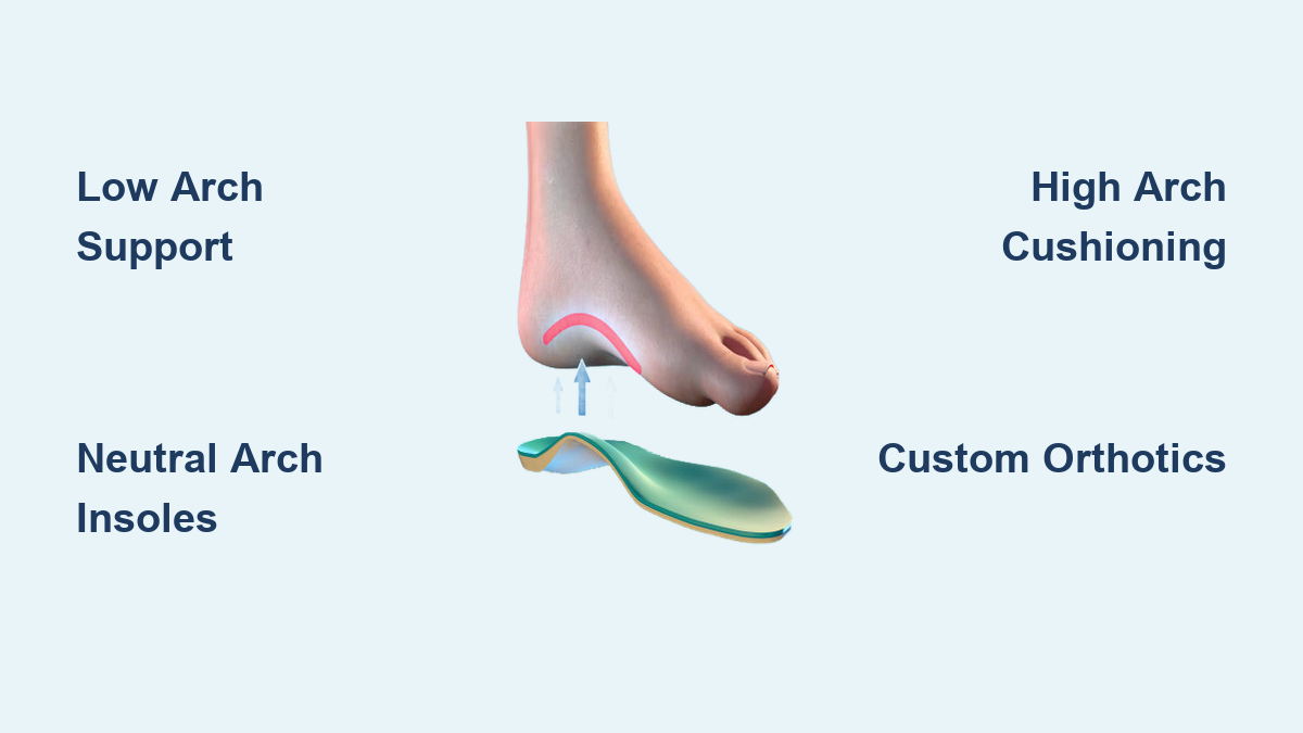
The medial longitudinal arch, that curved structure along the inside of your foot, plays a crucial role in shock absorption and weight distribution. But over time, the very tissues that hold it up weaken. Studies show 13% to 30% of adults have flat feet, with a significant number developing the condition later in life due to aging, injury, or chronic illness. Unlike congenital flat feet, age-related arch collapse often comes with pain, altered gait, and even knee or back discomfort.
In this guide, you’ll learn the real reasons behind why arch height changes as we get older, how to spot early signs, and what you can do—both preventively and therapeutically—to maintain foot function and comfort. From tendon degeneration to hormonal shifts and systemic diseases, we’ll break down the science into actionable insights.
Posterior Tibial Tendon Degeneration and Arch Collapse

The posterior tibial tendon (PTT) is the primary dynamic support for the medial arch. Running from the calf behind the inner ankle and attaching to bones in the midfoot, this tendon actively lifts and stabilizes the arch during walking. With age, however, it becomes vulnerable to wear, inflammation, and eventual failure—leading to posterior tibial tendon dysfunction (PTTD), the leading cause of adult-acquired flatfoot.
PTTD progresses in four distinct stages:
– Stage I: Inflammation and microtears cause pain along the inner ankle, but the arch still appears normal.
– Stage II: The tendon stretches or partially tears, allowing the arch to visibly drop and the heel to tilt outward (heel valgus).
– Stage III: Joints stiffen, and the deformity becomes fixed—even when not bearing weight.
– Stage IV: The ankle joint becomes involved, altering overall leg alignment.
Women over 40, individuals with diabetes, hypertension, or excess body weight are at higher risk. Repetitive stress from prolonged standing or high-impact activities accelerates damage. Without intervention, PTTD leads to irreversible structural change.
Pro Tip: If you feel persistent pain behind the inner ankle—especially when walking or climbing stairs—don’t dismiss it. This could be the earliest warning sign of arch collapse.
Spring Ligament Failure and Loss of Static Support
While tendons provide active, movement-based support, ligaments offer passive, structural stability. The spring ligament complex, located beneath the talus bone, acts as a sling that holds the arch in place. With aging, collagen in these ligaments deteriorates, reducing elasticity and leading to permanent stretching.
When both the posterior tibial tendon and spring ligament weaken, the arch loses both dynamic and static support. This dual failure is often the tipping point for visible flatfoot deformity. Unlike tendon issues, ligament damage is less likely to cause early pain—meaning the foot can collapse silently until significant deformity occurs.
Visual Clue: If your foot appears flatter in photos over time or you notice your heel leaning outward, it may signal ligamentous failure.
Hormonal Shifts and Pregnancy-Induced Arch Changes
Although not a direct aging factor, pregnancy can initiate long-term arch changes that become more apparent later in life. During pregnancy, the hormone relaxin increases to loosen pelvic ligaments for childbirth—but it affects connective tissue throughout the body, including the feet.
Combined with added body weight, this causes temporary flattening and widening of the foot. While some women regain their original foot shape postpartum, 60–70% permanently increase by half a shoe size or more. This early arch reduction sets the foundation for progressive collapse as other age-related factors accumulate.
Key Insight: Pregnancy can be a “first wave” of arch degeneration. If your feet changed during pregnancy, proactive foot care in midlife is essential to prevent further decline.
How Foot Injuries Trigger Progressive Arch Collapse

Even a single injury can destabilize the arch and lead to flatfoot years later. Tendon ruptures, ligament tears, or midfoot fractures disrupt the foot’s structural integrity.
For example:
– A ruptured posterior tibial tendon can cause immediate arch drop.
– A Lisfranc injury (midfoot ligament tear) alters tarsal alignment, leading to chronic flattening.
– Navicular or calcaneus fractures compromise key arch-supporting bones.
Even after healing, scar tissue and altered biomechanics prevent full recovery. If rehabilitation is incomplete or high-impact activity resumes too soon, the foot may slowly deform over time.
Warning Sign: If you’ve had a foot or ankle injury and now notice uneven shoe wear or inward rolling, get it evaluated. Early orthotic support can halt progression.
Systemic Diseases That Accelerate Arch Collapse
Certain medical conditions dramatically speed up arch degeneration by damaging nerves, joints, or connective tissue.
Diabetes and Charcot Foot
Diabetes contributes through peripheral neuropathy, which reduces sensation. Without pain feedback, micro-injuries go unnoticed, leading to abnormal loading and structural breakdown.
In severe cases, Charcot neuroarthropathy develops, causing:
– Bone resorption
– Joint dislocation
– Midfoot fractures
– A “rocker-bottom” foot appearance
Early signs include warmth, redness, and swelling—often without pain. If untreated, the arch collapses permanently.
Action Step: Diabetics should inspect their feet daily and avoid walking barefoot. Annual podiatric exams are critical.
Rheumatoid Arthritis (RA)
RA attacks synovial joints, including those in the midfoot. Chronic inflammation leads to:
– Cartilage and bone erosion
– Ligament laxity
– Tarsal bone subluxation
– Progressive arch flattening
Unlike PTTD, RA causes diffuse joint destruction, resulting in a more generalized collapse.
Visual Clue: RA patients often develop a “splay foot” with prominent midfoot bones and difficulty fitting shoes.
Obesity and Mechanical Overload
Excess body weight increases ground reaction forces on the foot by 3–4 times body weight during walking. Over decades, this sustained load fatigues tendons and stretches ligaments.
Obesity is a major risk factor for:
– PTTD
– Plantar fasciitis
– Early osteoarthritis
Even without other issues, carrying extra weight accelerates age-related arch decline.
Takeaway: Losing 10–15 pounds can significantly reduce stress on foot structures and slow arch collapse.
How Flattened Arches Change Foot Shape
As the arch lowers, the entire foot expands—lengthwise and widthwise.
Increased Foot Length and Width
When the arch flattens, the distance between heel and forefoot increases. Simultaneously, ligament laxity allows the tarsal and metatarsal bones to splay outward. This results in:
– Longer feet: Due to arch elongation
– Wider feet: Due to metatarsal spreading
– Shoe size increase: Often by half a size or more
Many older adults unknowingly wear shoes that are too narrow or short, increasing pressure points and ulcer risk—especially in diabetics.
Shoe Fit Challenges
Standard shoe widths don’t accommodate age-related widening. Look for:
– Wide or extra-wide options
– Stretchable uppers
– Adjustable closures (e.g., Velcro)
Pro Tip: Get your feet measured every 2–3 years. Most people are surprised to find they’ve grown.
Gait and Posture Consequences of Flat Feet
A collapsed arch doesn’t just affect the foot—it alters your entire movement pattern.
Excessive Pronation and Kinetic Chain Effects
Flat feet lead to overpronation, where the foot rolls inward excessively during walking. This misalignment travels up the body:
– Ankles: Increased strain on medial ligaments
– Knees: Internal rotation and valgus stress, raising IT band and patellofemoral pain risk
– Hips: Altered pelvic tilt and gluteal weakness
– Lower back: Compensatory hyperlordosis and disc pressure
Result: You may develop knee, hip, or back pain—without realizing the root cause is your feet.
Reduced Shock Absorption
The arch acts like a spring, storing and releasing energy with each step. A collapsed arch loses this elastic function, transferring more impact to joints. This contributes to:
– Early osteoarthritis
– Joint fatigue
– Generalized leg tiredness
Fact: The average person walks 75,000–115,000 miles in a lifetime—equivalent to 3+ trips around Earth. Your feet need to be resilient.
Early Warning Signs of Arch Collapse
Not all flat feet hurt, but when symptoms appear, they often progress.
Common Symptoms to Watch For
- Pain along the inner foot or ankle
- Swelling on the inner ankle
- Difficulty standing on one toe
- Tired, aching feet by day’s end
- Outward-pointing toes due to heel valgus
- Leg cramps after walking
Symptoms typically worsen with activity and improve with rest. In advanced stages, pain may shift to the outer foot due to joint impingement.
Red Flag: If you can’t rise onto your toes on one foot, see a foot specialist. This is a strong indicator of PTTD.
How Diagnosis Works
Early detection improves outcomes. Diagnosis combines clinical tests and imaging.
Key Diagnostic Tests
- Single-leg heel rise test: Inability to rise on one foot suggests PTTD.
- Too many toes sign: When viewed from behind, more lateral toes are visible due to heel tilt.
- Flexible vs. rigid flatfoot: If the arch reappears when standing on tiptoes, it’s flexible and potentially correctable.
Imaging for Confirmation
- Weight-bearing X-rays: Show arch angle, heel alignment, and joint space narrowing.
- MRI: Evaluates tendon tears (especially PTT) and soft tissue damage.
- CT scans: Used for surgical planning in complex cases.
Note: Imaging is crucial before surgery but often unnecessary for early-stage management.
Non-Surgical Treatment Options
Most cases improve with conservative care—especially when caught early.
Custom Orthotics for Arch Support
Custom-made orthotics are the gold standard. They:
– Support the medial arch
– Align the heel
– Reduce strain on the posterior tibial tendon
– Improve gait efficiency
Made from a cast or 3D scan, modern orthotics fit into most shoes—including dress styles.
Evidence: Studies show orthotics reduce pain and improve function in 70–80% of PTTD patients.
Supportive Footwear Essentials
Choose shoes with:
– Stiff soles
– Firm heel counters
– Built-in arch support
– Wide toe boxes
Avoid flip-flops and high heels—they increase arch strain.
Replacement Rule: Change shoes every 300–500 miles or when soles show wear.
Strengthening and Stretching Exercises
Daily exercises can slow or reverse early collapse.
Key Exercises:
- Towel curls: Scrunch a towel with your toes (3 sets of 15)
- Marble pickups: Use toes to move marbles into a cup
- Calf stretches: Hold 30 seconds, 2–3 times daily
- Heel raises: Stand on one foot and rise onto toes (build up to 3 sets of 10)
Time Commitment: Just 5–10 minutes daily can make a difference.
When Surgery Is Needed
Surgery is considered when conservative treatments fail and pain or deformity limits function.
Indications for Surgery
- Rigid, painful flatfoot
- Failed orthotics and physical therapy
- Severe arthritis
- Charcot foot instability or ulcer risk
Common Procedures
- Tendon transfer: Replace damaged PTT with another tendon
- Osteotomy: Cut and realign bone to restore arch height
- Arthrodesis: Fuse joints to stabilize the foot
- Lateral column lengthening: Rebalance foot structure
Recovery: Typically 3–6 months, with immobilization and gradual return to activity.
Outcome: Most patients report improved alignment, reduced pain, and better mobility.
Preventive Foot Care for Aging Adults
You can’t stop aging—but you can slow arch decline with smart habits.
Regular Foot Assessments
- See a podiatrist annually, especially if you have diabetes or neuropathy.
- Get feet measured regularly—shoe size changes are normal.
- Inspect feet daily for cuts, calluses, or swelling.
Daily Foot Maintenance
- Moisturize nightly to prevent cracking.
- Wear cotton socks to reduce friction.
- Never walk barefoot—even at home.
Lifestyle Adjustments
- Maintain a healthy weight to reduce foot load.
- Stay active with low-impact exercise.
- Incorporate balance and flexibility training to prevent falls.
Final Tip: Your feet carry you through life. Treat them well, and they’ll carry you farther.