Pain on the inner side of the midfoot, right at the top of the arch, is easy to ignore at first. It may begin as a dull ache after a run, a long day on your feet, or a new pair of shoes. Over time, though, it can become sharp, persistent and limiting. In many of these cases, the culprit is a small but very important bone called the navicular.
This guide explains what navicular bone pain is, why it happens, how it is diagnosed, and which treatment and recovery options are commonly used. It also covers footwear, home care, prevention and the warning signs that mean you should see a healthcare professional without delay.
Quick Summary
- The navicular is a boat-shaped bone on the inner side of the midfoot that helps support the arch.
- Pain is most often linked to overuse, stress fractures, an accessory navicular, tendon problems and flat feet.
- Early rest, supportive footwear and proper diagnosis improve outcomes significantly.
- Persistent midfoot pain, especially in athletes, should be examined and imaged rather than simply ignored.
Table of Contents
- What Is the Navicular Bone?
- Why the Navicular Bone Is Prone to Pain
- Common Causes of Navicular Pain
- Who Is at Higher Risk?
- Symptoms to Watch For
- How Navicular Pain Is Diagnosed
- Treatment Options
- Home Care and Self-Management
- Choosing Footwear and Arch Support
- Gentle Exercises for Recovery
- Recovery Timeline
- Prevention Tips
- When to See a Doctor
- Frequently Asked Questions
- Final Thoughts
What Is the Navicular Bone?
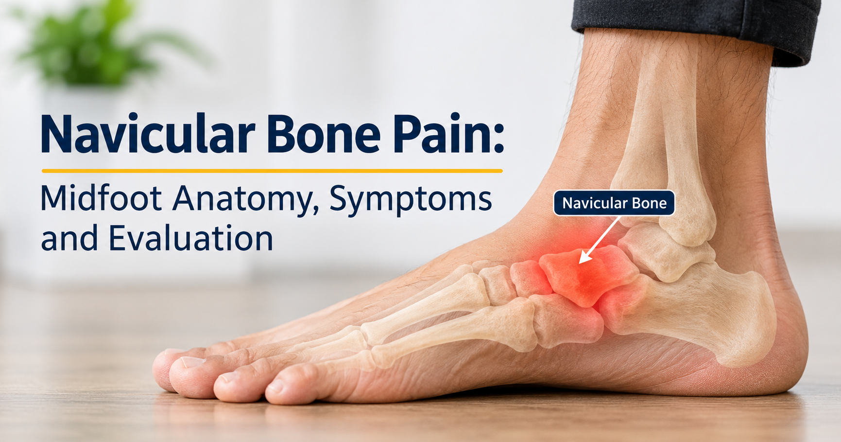
The navicular is one of the seven tarsal bones of the foot. Its name comes from the Latin word for “little boat”, which describes its curved shape. It sits on the inner (medial) side of the midfoot, between the talus (ankle bone) behind it and the three cuneiform bones in front of it.
You can often feel a small bony bump on the inner side of your foot, a little in front of the ankle and slightly above the arch. That prominence is the navicular, or its tuberosity, which is the attachment point for an important tendon called the tibialis posterior.

What Does the Navicular Do?
- Supports the medial arch: It acts like the keystone of a stone arch, helping the arch hold its shape.
- Transfers load: It passes forces from the ankle to the forefoot during walking, running and jumping.
- Anchors the tibialis posterior tendon: This tendon helps lift the arch and stabilize the foot with every step.
- Absorbs shock: Together with the surrounding joints, it helps cushion the impact of landing.
Why the Navicular Bone Is Prone to Pain
The navicular is a high-stress location. Forces from the ankle meet forces from the forefoot at this single bone, and it must handle them thousands of times a day. Several features make it vulnerable:
- Heavy load concentration: The central part of the bone is compressed between the talus and the cuneiforms during push-off.
- Limited blood supply: The central third of the bone receives relatively less blood flow than other areas, which can slow healing when a stress injury occurs.
- Tendon attachment: The pull of the tibialis posterior tendon can irritate the bone or its attachment site.
- Position in the arch: Anything that changes arch mechanics, such as flat feet or high arches, changes the stress on the navicular.
Common Causes of Navicular Pain
Navicular pain is not one single condition. It is a symptom that can come from several different problems. Understanding the most common causes helps you discuss the issue clearly with a clinician.

1. Overuse and Repetitive Stress
Running, sprinting, jumping and court sports repeatedly load the midfoot. If training volume or intensity rises faster than the bone can adapt, the area becomes irritated and sore. This is often the earliest stage, before an actual crack develops.
2. Navicular Stress Fracture
A stress fracture is a small crack caused by repeated loading rather than a single injury. It is more common in athletes involved in track, sprinting, basketball, soccer, gymnastics and dance. Because of the bone’s limited blood supply, these fractures can be slow to heal and are sometimes difficult to see on a basic X-ray. The pain is often vague at first, felt in the top of the arch, and gets worse with push-off or jumping.
3. Accessory Navicular Syndrome
Some people are born with an extra piece of bone or cartilage next to the navicular, known as an accessory navicular. Many people never notice it. In others, it forms a visible bump on the inner foot that rubs against shoes or pulls on the tibialis posterior tendon, causing tenderness, redness and pain, especially during adolescence or after a twist or sports strain.
4. Posterior Tibial Tendon Problems
The tibialis posterior tendon attaches on or near the navicular. When this tendon is overworked, inflamed or degenerated, pain can be felt along the inner ankle and at the navicular attachment. Over time, a weakened tendon may allow the arch to flatten, which in turn loads the navicular even more.
5. Flat Feet or Excessive Pronation
When the arch collapses excessively during standing or walking, the navicular tends to drop and may press against the inside of the shoe. This changes joint mechanics and can create ongoing pain in the arch and midfoot.
6. Poor or Unsupportive Footwear
Thin, flexible or worn-out shoes provide little midfoot support. Tight shoes can also press directly on the bony bump on the inner foot. Both situations can irritate the region over time.
7. Less Common Conditions
- Köhler disease: A temporary condition in young children, usually between ages 4 and 7, in which blood supply to the developing navicular is briefly reduced, causing a limp and midfoot pain.
- Müller-Weiss syndrome: A rare condition, mostly seen in adults, in which the navicular gradually changes shape or collapses, leading to chronic midfoot pain.
- Avascular necrosis: Loss of blood supply that can weaken the bone, sometimes following a fracture.
- Arthritis of the midfoot: Wear of the joints surrounding the navicular, producing stiffness and aching.
- Direct injury: A fall, twisting injury or heavy blow to the midfoot can bruise or fracture the bone.
Who Is at Higher Risk?
Anyone can develop navicular pain, but certain groups see it more often:
- Runners, sprinters, jumpers, soccer and basketball players
- Dancers and gymnasts
- People who suddenly increase training intensity, mileage or hard-surface activity
- People with flat feet, very high arches or limited ankle flexibility
- Adolescents with an accessory navicular
- Individuals who stand or walk on hard floors for long hours
- People wearing worn-out or unsupportive shoes
- Those with lower bone density or nutritional deficiencies affecting bone health
Symptoms to Watch For
Symptoms vary depending on the underlying cause, but several signs appear repeatedly in people with navicular-area problems.

- Dull ache on the top or inner side of the arch, often vague and hard to pinpoint at first.
- Pain that increases with activity, especially running, jumping, climbing stairs or pushing off the toes.
- Tenderness to touch over the bony prominence on the inner midfoot.
- Mild swelling or redness around the inner arch, particularly with an accessory navicular.
- Pain that lingers after exercise or aches at rest or at night in more advanced cases.
- Changes in walking pattern, such as walking on the outer edge of the foot to avoid pressure.
- Stiffness in the midfoot or reduced comfort in tight or narrow shoes.
Important
Pain that keeps returning at the same spot during activity and eases with rest should not be dismissed as simple soreness. Early assessment can prevent a minor stress reaction from becoming a full fracture.
How Navicular Pain Is Diagnosed
A proper diagnosis begins with a conversation and a hands-on examination. A clinician will usually ask when the pain began, what activities make it worse, whether your training or footwear changed recently, and whether you have had previous foot injuries.
Physical Examination
- Pressing on the navicular (often called the “N spot”) to check for tenderness
- Assessing arch shape and foot alignment while standing and walking
- Testing the strength and pain response of the tibialis posterior tendon
- Checking ankle and midfoot range of motion
- Observing single-leg hopping or toe-rise movements if appropriate
Imaging Tests
| Test | What It Shows |
|---|---|
| X-ray | Bone shape, an accessory navicular, obvious fractures, arthritis; may miss early stress fractures |
| MRI | Bone stress reactions, early stress fractures, tendon problems and soft tissue inflammation |
| CT scan | Detailed view of fracture lines, healing progress and bone structure |
| Bone scan | Highlights areas of increased bone activity when the source of pain is unclear |
| Ultrasound | Assesses the tibialis posterior tendon and surrounding soft tissues |
Treatment Options
Treatment depends on the cause, severity and your activity level. The overall goal is to calm the pain, protect the bone, restore normal foot mechanics and return you safely to your usual activities.

Non-Surgical Treatment
Most navicular problems improve without surgery when they are identified early.
- Activity modification: Reduce or stop the activity that triggers pain. Low-impact alternatives such as swimming or upper-body training may be suitable.
- Protected weight-bearing: For stress fractures, clinicians commonly recommend a walking boot, a cast or crutches to keep load off the bone for several weeks.
- Ice therapy: Applying a cold pack wrapped in a thin cloth for 15–20 minutes can help ease soreness and swelling.
- Anti-inflammatory medicines: Some people use over-the-counter pain relievers, but you should ask a pharmacist or doctor first, particularly if a fracture is suspected, since medication choices may matter for bone healing.
- Orthotic support: Custom or prefabricated insoles can reduce strain on the arch and tendon.
- Physical therapy: Strengthening, stretching and gait retraining address muscle imbalance and mechanics.
- Footwear changes: Roomier, more supportive shoes reduce direct pressure on the inner bump.
Surgical Treatment
Surgery is usually considered only when conservative care does not work or when the problem is more serious. Examples include:
- A stress fracture that fails to heal, or a displaced fracture
- A painful accessory navicular that continues to cause symptoms despite proper non-surgical care
- Advanced collapse or deformity of the bone
- Significant tendon damage or severe flatfoot deformity
Procedures may involve fixing the fracture with screws, removing the extra bone and reattaching the tendon, or in selected cases fusing joints. Your surgeon will explain which approach suits your situation and what recovery will involve.
Home Care and Self-Management
While waiting for an appointment or during recovery, these steps may help keep you comfortable. They do not replace professional assessment, particularly if a fracture is possible.
- Rest the foot: Avoid running, jumping and long walks until you know the cause.
- Elevate when seated: Raising the foot above hip level can help with swelling.
- Use cold therapy: Short, regular sessions help many people, but never place ice directly on bare skin.
- Wear supportive shoes indoors: Walking barefoot on hard floors can aggravate arch pain.
- Add cushioning: A soft pad or sleeve over the inner foot bump can reduce rubbing.
- Keep moving gently: Within pain limits, maintain overall fitness with non-impact exercise recommended by your clinician.
- Track your symptoms: Note which activities hurt, how long pain lasts and whether it is improving or worsening.
Choosing Footwear and Arch Support
Footwear is one of the most practical factors you can control. The right pair can reduce stress on the navicular, while the wrong pair can keep the problem going.
What to Look for in Shoes
- Firm heel counter: Helps stabilize the rear foot and limit excessive inward rolling.
- Good midfoot structure: The shoe should resist excessive twisting through the middle.
- Appropriate arch contour: Support should match your arch type rather than pushing too aggressively.
- Cushioned midsole: Reduces repetitive impact loading.
- Adequate width and soft upper: Prevents pressure on the inner foot bump.
- Removable insole: Allows you to fit a custom or supportive insert if needed.
Types of Insoles and Supports
| Support Type | Best For | Notes |
|---|---|---|
| Cushioned comfort insoles | Mild aching and long hours on the feet | Add shock absorption but limited structural control |
| Semi-rigid arch supports | Flat feet, overpronation, tendon strain | Help control arch collapse while remaining comfortable |
| Custom orthotics | Persistent pain, structural problems | Made from a foot assessment; usually the highest level of tailoring |
| Padded accessory-bump sleeves | Rubbing over an accessory navicular | Reduce friction and pressure on the inner foot |
If you use a new insole, break it in gradually over several days. A good support should feel stable and comfortable, not sharp or painful.
Gentle Exercises for Recovery
Do not begin strengthening exercises while a fracture is suspected or unconfirmed. Once your clinician confirms it is safe, a gradual program may include the following. Stop any exercise that causes sharp or increasing pain.
- Toe curls with a towel: Scrunch a towel with your toes to wake up the small muscles under the arch.
- Short foot exercise: Gently draw the ball of the foot toward the heel without curling the toes, lifting the arch slightly.
- Calf stretches: Tight calves increase midfoot stress, so stretch gently against a wall for 30 seconds, repeating a few times.
- Heel raises: Rise slowly onto the balls of both feet, then lower with control, progressing to single-leg raises as pain allows.
- Resistance band inversion: Turn the foot inward against light band resistance to strengthen the tibialis posterior.
- Balance training: Standing on one leg improves stability and foot control.
Hip and core strengthening also helps, since better control higher up the leg reduces abnormal stress through the foot.
Recovery Timeline
Healing times vary widely, and your own timeline will depend on the diagnosis, your age, bone health and how closely you follow your plan.
| Condition | Typical Recovery Pattern |
|---|---|
| Overuse irritation | Often several weeks of reduced load and supportive footwear |
| Stress fracture (non-surgical) | Commonly around 6–8 weeks of protected loading, followed by a gradual return to sport over additional weeks |
| Accessory navicular syndrome | Weeks to a few months with supports; longer if surgery is needed |
| Tendon-related pain | Several weeks to months with rehabilitation |
| Surgical cases | Varies by procedure; often several months before full activity |
Returning too soon is one of the most common reasons navicular problems come back. Progress in stages, and increase activity only when walking and daily tasks are pain-free.
Prevention Tips
- Increase training gradually: Raise distance, speed or jumping volume in small steps rather than sudden jumps.
- Replace worn shoes: Running shoes lose cushioning and support over time.
- Vary surfaces and activities: Cross-training reduces repeated stress on one area.
- Strengthen the foot and lower leg: Strong calves, arch muscles and the tibialis posterior help protect the navicular.
- Address flat feet early: Appropriate supports can lessen long-term strain.
- Support bone health: Eat a balanced diet with enough calcium, vitamin D and protein, and discuss supplements with a professional if you suspect a deficiency.
- Listen to early pain: A small ache that keeps returning in the same spot is a signal to rest and get checked.
- Allow recovery days: Bone adapts during rest, not only during training.
When to See a Doctor
Seek medical advice promptly if you notice any of the following:
- Pain that persists for more than a week or two despite rest
- Pain at the top or inner side of the arch that worsens with activity
- Pain after a fall, twist or direct impact to the foot
- Difficulty putting weight on the foot or a limp that does not improve
- Visible swelling, bruising or deformity
- Pain at night or at rest
- Numbness, tingling, warmth, redness spreading or fever
- Midfoot pain in a young child with a limp
A podiatrist, sports medicine doctor or orthopedic foot specialist is usually best placed to evaluate navicular problems.
Frequently Asked Questions
Where exactly is navicular pain felt?
Typically on the inner side of the midfoot, near the top of the arch, slightly in front of the ankle. Some people describe it as a deep ache in the arch rather than a precise spot.
Can I keep walking with navicular pain?
Light daily walking may be tolerable in mild overuse cases, but if a stress fracture is possible you should limit weight-bearing and get assessed. Continuing to load a cracked bone can delay healing or worsen the injury.
Does a normal X-ray rule out a navicular stress fracture?
Not always. Early stress fractures of the navicular often do not appear on plain X-rays. If symptoms strongly suggest one, a clinician may recommend an MRI or CT scan.
Is an accessory navicular dangerous?
No. It is a common anatomical variation, and many people live with it without symptoms. It only needs treatment if it becomes painful or interferes with activity.
Can flat feet cause navicular pain?
Yes. When the arch drops excessively, the navicular may bear more stress or press against the shoe, and the tibialis posterior tendon has to work harder. Supports and strengthening often help.
How long does a navicular stress fracture take to heal?
It commonly takes around two months or longer of protected loading, with a further gradual return to sport. Healing can be slower than in other bones because of the area’s limited blood supply, so following professional guidance is essential.
Will I need surgery?
Many cases settle with rest, protection, supports and rehabilitation. Surgery is generally reserved for fractures that do not heal, displaced fractures, persistent accessory navicular pain or significant structural problems.
Can I run again after navicular pain?
Most people can return to running after proper healing and rehabilitation. The key is a gradual, pain-free progression and addressing the underlying cause, such as training errors, footwear or foot mechanics.
Final Thoughts
Navicular bone pain may start quietly, but this small bone plays a large role in how your foot supports and moves your body. Whether the cause is overuse, a stress fracture, an accessory navicular, tendon strain or flat feet, early attention makes a real difference. Rest the area, wear supportive shoes, use appropriate arch support, and seek an accurate diagnosis when pain does not settle.
With the right care, patience and a gradual return to activity, most people recover well and get back to the things they enjoy while protecting their feet for the long run.
Medical Disclaimer
This article is for general educational purposes only and is not a substitute for professional medical advice, diagnosis or treatment. Always consult a qualified healthcare provider about foot pain, suspected fractures or before starting any new treatment or exercise program.