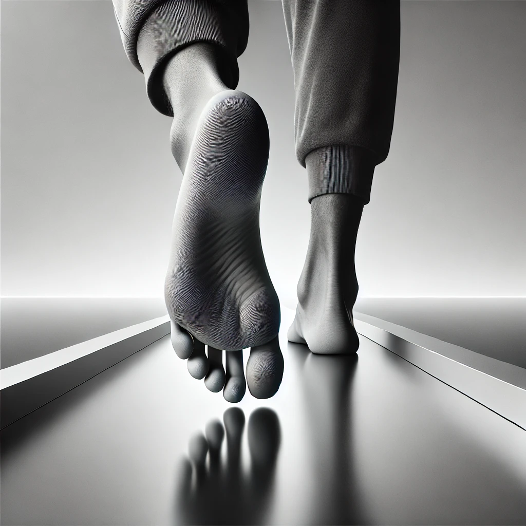
If you’ve ever pushed off the ground while sprinting, stood on tiptoes, or simply walked barefoot on a hard surface, your metatarsal bones were hard at work. These five long bones in the midfoot are central to how we move, balance, and absorb impact with every step. Though often overlooked, the metatarsal bones are critical structural components that link the rearfoot to the toes, transforming the foot from a flexible adapter into a rigid lever when needed.
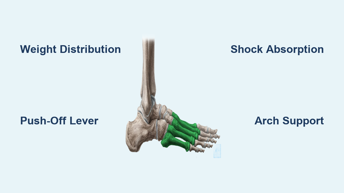
Each foot contains five metatarsals—numbered I to V, starting from the big toe (medial side) to the little toe (lateral side). They connect the tarsal bones of the ankle and midfoot to the phalanges, or toe bones, forming the metatarsus—a dynamic framework essential for weight transfer, propulsion, and stability. The first metatarsal, attached to the big toe, is the shortest but thickest and strongest, bearing up to 30% of body weight during walking and even more during running. Meanwhile, the second metatarsal is the longest, making it particularly vulnerable to stress fractures in athletes.
Understanding what the metatarsal bone is and its function goes beyond anatomy—it’s key to preventing and managing common foot problems like metatarsalgia, gout, bunions, and stress fractures. Whether you’re an athlete, someone on your feet all day, or just looking to maintain mobility, knowing how these bones work helps you recognize pain signals early and take protective action. In this guide, we’ll explore their location, structure, biomechanical roles, and the most common conditions that affect them.
Where Are the Metatarsal Bones Located?
Five Long Bones Spanning the Midfoot
The metatarsal bones run through the midsection of the foot, forming a bridge between the tarsals (like the cuboid and cuneiforms) at the back and the proximal phalanges at the front. Each metatarsal corresponds to a toe:
- Metatarsal I: Connects to the big toe (hallux)
- Metatarsal II: Second toe
- Metatarsal III: Middle toe
- Metatarsal IV: Fourth toe
- Metatarsal V: Little toe
This arrangement creates the metatarsal parabola, a curved alignment where the second metatarsal typically extends the furthest forward. This shape helps distribute pressure evenly across the ball of the foot during standing and walking. When this balance is disrupted—such as when one metatarsal is longer or lower than the others—it can lead to localized pain and calluses.
How to Identify Metatarsal Position
You can feel the heads of the metatarsals on the sole of your foot just behind the toes, especially when standing. The first, second, and fifth metatarsal heads are the most prominent and bear the brunt of weight during push-off. Pain under the second or third metatarsal head often signals metatarsalgia, while a sharp ache near the base of the fifth may indicate a Jones fracture. Knowing the location of each bone helps pinpoint the source of discomfort and guides proper treatment.
Anatomy of a Metatarsal Bone: Base, Shaft, and Head
Three Structural Regions for Strength and Flexibility
Each metatarsal bone has three distinct parts:
- Base (proximal end): The rear portion that articulates with the tarsal bones. It’s wedge-shaped and varies in size, with the first metatarsal base being the largest.
- Shaft (body): The long, slender middle section. It curves slightly upward (dorsally convex), contributing to the foot’s natural arches.
- Head (distal end): The rounded front end that forms the metatarsophalangeal (MTP) joint with the toe bones.
This design allows the metatarsals to be both strong and flexible. The dorsal convexity of the shaft provides space for tendons to glide over the top of the foot, while the tapered shape enables efficient force transmission from heel to toe.
Why Shape Influences Foot Health
The rounded heads of the metatarsals press into the ground during weight-bearing, making them natural pressure points. If one metatarsal is plantarflexed (dropped too low), it can overload that area, leading to pain, inflammation, or callus formation. Similarly, a short first metatarsal shifts excess load to the second, increasing fracture risk. The longitudinal curvature also supports the medial arch; if this curve flattens due to flat feet or ligament laxity, it strains the metatarsals and surrounding tissues.
How Metatarsals Connect to Other Foot Bones

Tarsometatarsal (Lisfranc) Joints for Midfoot Stability
The bases of the metatarsals connect to the tarsal bones at the tarsometatarsal (TMT) joints, also known as Lisfranc joints:
- Metatarsal I → Medial cuneiform
- Metatarsal II → Intermediate cuneiform (acts as a central keystone)
- Metatarsal III → Lateral cuneiform
- Metatarsals IV and V → Cuboid
These joints are stabilized by strong ligaments, especially the Lisfranc ligament between the medial cuneiform and second metatarsal. Injuries here—like a Lisfranc fracture-dislocation—can severely disrupt foot mechanics and often require surgery.
Metatarsophalangeal (MTP) Joints Enable Toe Motion
At the front, each metatarsal head forms an MTP joint with the corresponding toe. These joints allow:
- Flexion and extension (toe bending)
- Abduction and adduction (toe spreading)
The first MTP joint is the most critical for propulsion, handling forces up to three times body weight during running. Limited motion here—due to arthritis, bunions, or tight shoes—can impair gait and increase stress on other metatarsals.
Intermetatarsal Joints Allow Subtle Adjustments
Adjacent metatarsal bases also connect via intermetatarsal joints, stabilized by interosseous ligaments. These small joints allow slight gliding motions, helping the foot adapt to uneven surfaces while maintaining structural integrity.
Muscles That Control Metatarsal Position

Key Muscles Attached to the Metatarsals
While muscles don’t move the metatarsals directly, they stabilize and influence their position during movement:
| Metatarsal | Muscle | Role |
|---|---|---|
| I | Tibialis anterior | Dorsiflexes foot, supports arch |
| Fibularis longus | Plantarflexes first ray, supports transverse arch | |
| 1st dorsal interosseous | Abducts big toe, stabilizes MTP joint | |
| II–V | Dorsal and plantar interossei | Move toes side-to-side, stabilize MTP joints |
| V | Fibularis brevis | Everts foot, stabilizes lateral column |
| Flexor digiti minimi brevis | Flexes little toe |
The fibularis longus tendon runs beneath the foot and attaches to the base of the first metatarsal, acting like a sling that supports both the transverse and medial arches.
Role of the Interossei Muscles
The dorsal interossei (four muscles) abduct the toes (spread them outward), while the plantar interossei (three muscles) adduct them (pull them inward). Together, they maintain toe alignment and prevent clawing or splaying. Weakness or imbalance can lead to deformities like Morton’s toe or hammer toes, altering pressure distribution and increasing metatarsal stress.
Ligaments That Stabilize the Metatarsals
Deep Transverse Metatarsal Ligaments (DTML)
Four fibrous bands connect the heads of adjacent metatarsals just behind the MTP joints. These DTMLs prevent toe splaying and help maintain the transverse arch. They integrate with the plantar plates—thickened ligaments on the bottom of the MTP joints—that resist hyperextension and vertical loading.
In bunions (hallux valgus), these ligaments stretch or tear, allowing the first metatarsal to drift medially and the big toe to shift laterally.
Plantar Ligaments for Joint Support
The plantar ligaments reinforce the bottom of the MTP joints, guide flexor tendons, and act as checkreins against excessive dorsiflexion during push-off. Damage—such as in capsulitis or dislocation—can cause instability and chronic pain.
Interosseous Ligaments for Midfoot Rigidity
Located between the metatarsal bases, interosseous ligaments bind the bones together and resist separation during weight-bearing. Along with dorsal and plantar ligaments, they form a strong network that maintains midfoot stability during walking.
How Metatarsals Support the Foot’s Arch System

Medial Longitudinal Arch: Primary Shock Absorber
The first, second, and third metatarsals form the front of the medial longitudinal arch, the highest and most shock-absorbent arch. Supported by the tibialis posterior tendon, spring ligament, and plantar fascia, this arch collapses if the first metatarsal drops, leading to metatarsalgia or flatfoot.
Lateral Longitudinal Arch: Stability During Push-Off
The fourth and fifth metatarsals support the lateral arch, which is lower and stiffer. This arch provides balance during midstance and is stabilized by the fibularis longus and brevis tendons, creating a “tie-beam” effect across the foot.
Transverse Arch: Prevents Forefoot Splaying
The bases of all five metatarsals form the transverse arch, which resists compression. Key supports include the deep transverse metatarsal ligaments and the peroneus longus tendon. Loss of this arch leads to forefoot splaying, common in aging or connective tissue disorders.
What Do Metatarsal Bones Do? Core Functions Explained
Weight-Bearing: Distributing Body Load
During walking, weight shifts from heel to forefoot. As the heel lifts, load transfers to the metatarsal heads, which press into the ground. The first metatarsal head carries about 30% of forefoot load, while the second and third share the rest. Proper alignment ensures even distribution.
Propulsion: The Push-Off Phase
During toe-off, the metatarsals act as rigid levers. The windlass mechanism—where toe extension tightens the plantar fascia—raises the arch and stiffens the foot for efficient push-off. Restricted MTP motion disrupts this, reducing propulsion and increasing joint stress.
Shock Absorption and Balance
While rigid during push-off, the metatarsals allow slight flexibility to absorb impact. Combined with soft tissue padding and arch elasticity, they cushion landing forces. On uneven terrain, subtle joint motions help maintain balance and prevent falls.
Common Metatarsal Injuries and Conditions
Metatarsalgia: Forefoot Pain from Overload
Metatarsalgia causes burning or aching pain under the ball of the foot. Causes include high-impact activity, thin fat pads, or anatomical issues like a long second metatarsal. Treatment involves proper footwear, orthotics, and activity modification.
Stress Fractures: Tiny Cracks from Repetitive Stress
Common in runners, stress fractures occur in the second and third metatarsals. Symptoms include gradual pain that worsens with activity. Healing takes 6–8 weeks and may require immobilization.
Jones Fracture: High-Risk Break at the Fifth Metatarsal
A Jones fracture occurs at the base of the fifth metatarsal, where blood supply is poor. It often requires surgery, especially in athletes, and takes 8–12 weeks to heal.
Gout: Inflammation at the Big Toe Joint
Gout (podagra) most commonly affects the first MTP joint, causing sudden, severe pain, redness, and swelling. Uric acid crystals form due to cooler joint temperature. Repeated attacks can damage the metatarsal head.
Hallux Valgus: Bunion Deformity
In hallux valgus, the first metatarsal drifts inward, and the big toe angles outward, forming a bunion. This misalignment increases pressure on other metatarsals. Causes include genetics, narrow shoes, and ligament laxity.
How to Protect Your Metatarsal Bones
Wear Supportive, Well-Fitted Shoes
Choose shoes with:
– Wide toe box
– Low heel
– Cushioned sole
– Firm midsole
Avoid high heels and pointy shoes that compress the forefoot.
Use Protective Gear When Needed
In industrial jobs, metatarsal guards in boots protect against falling objects. Athletes can use silicone padding in socks to reduce fracture risk.
Prevent Overuse Injuries
- Increase activity gradually
- Replace worn shoes
- Strengthen foot muscles with towel curls and toe spreads
- Rest if you feel persistent forefoot pain
Final Note: The metatarsal bones are far more than passive connectors—they are dynamic, load-bearing structures essential for every step you take. Understanding what the metatarsal bone is and its function empowers you to recognize warning signs of injury and make informed choices about footwear, activity, and care. Whether you’re an athlete, someone on your feet all day, or just looking to maintain mobility, protecting your metatarsals ensures lasting foot health.