If you’ve ever taken your first step in the morning and felt a sharp, stabbing pain under your heel—like you’re walking on glass—you may be dealing with plantar fasciitis. It’s the most common cause of heel pain in adults, affecting millions each year. Despite its prevalence, many people don’t fully understand what it really is. So, what is plantar fasciitis exactly? It’s not just “inflammation” as the name suggests, nor is it simply a heel spur. Instead, it’s a degenerative condition of a key foot structure—the plantar fascia—caused by repetitive strain and biomechanical stress.
This condition impacts up to 10% of people in their lifetime, with peak onset between ages 40 and 60. While often linked to runners or those on their feet all day, it can affect anyone—especially those with tight calves, flat feet, high arches, or who wear unsupportive shoes. The hallmark sign? First-step pain: intense discomfort when getting out of bed or standing after rest. But beyond the pain, understanding the root causes, anatomy involved, and effective treatments is essential for recovery. In this guide, we’ll break down exactly what plantar fasciitis is—from its true pathology to how it develops, how it’s diagnosed, and what you can do to heal and prevent it for good.
The Plantar Fascia: Structure and Function

A Critical Support System in Your Foot
The plantar fascia is a thick, web-like band of connective tissue that runs along the sole of your foot, connecting your heel bone (calcaneus) to the base of your toes. Unlike a muscle or tendon, it’s a ligament—designed to support the longitudinal arch of the foot. Think of it like a bowstring under tension, maintaining the foot’s natural curve and acting as a shock absorber during walking and running.
This tissue bears significant mechanical load—up to 14 times your body weight during running—making it highly susceptible to overuse. It’s strongest at its origin: the medial calcaneal tubercle, the inner part of the heel bone. This is also the most common site of pain in plantar fasciitis.
How It Powers Your Step: The Windlass Mechanism
During walking, the plantar fascia plays a crucial role in foot stability and propulsion. As you push off with your big toe, the fascia tightens—a mechanism known as the windlass effect. This action lifts the arch, stiffens the foot, and converts it into a rigid lever for efficient forward movement.
When this mechanism is impaired—due to limited big toe motion or tight calf muscles—the plantar fascia is subjected to excessive tension. Over time, this leads to microtears, collagen breakdown, and degeneration, not classic inflammation. That’s why many experts now prefer the terms plantar fasciosis or plantar fasciopathy, which more accurately reflect its degenerative nature.
Why “Fasciitis” Is a Misleading Name
It’s Degeneration, Not Inflammation
Despite the name ending in -itis (which implies inflammation), modern research shows that inflammatory cells are rarely present in affected tissue. Instead, microscopic analysis reveals scarring, disorganized collagen, and fatty deposits—hallmarks of chronic degeneration. This shift in understanding has led to a reclassification of the condition as plantar fasciopathy, emphasizing wear-and-tear over acute inflammation.
Calling it “fasciitis” can mislead both patients and clinicians into thinking anti-inflammatory treatments alone will cure it—which they often don’t. The real issue? Repetitive mechanical overload leading to structural failure of the fascia.
How Microtears Lead to Chronic Pain
Every step you take puts stress on the plantar fascia. When the load exceeds the tissue’s capacity—due to poor biomechanics, sudden activity increases, or structural foot issues—tiny microtears form at the heel attachment. Normally, the body repairs these. But with ongoing strain, the repair process fails, leading to chronic degeneration.
Over time, the fascia becomes thicker, less elastic, and more painful, especially when stretched after periods of rest. This explains why pain is worst in the morning: the fascia has tightened overnight and is suddenly stretched upon weight-bearing.
Recognizing the Classic Symptoms
First-Step Heel Pain: The Telltale Sign
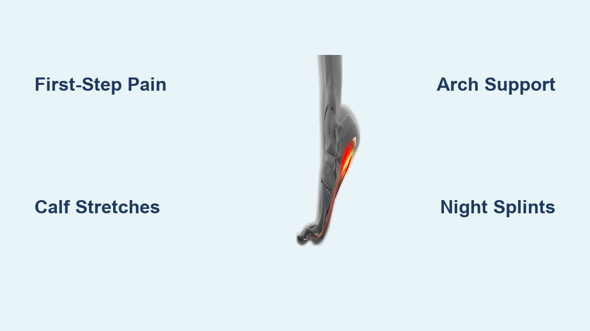
The #1 symptom of plantar fasciitis is sharp, stabbing pain under the heel with the first few steps after waking up or sitting. This “first-step pain” is so characteristic that it’s often diagnostic on its own. The pain typically lessens after a few minutes of walking as the fascia warms up—but may return after prolonged standing or activity.
Patients often describe it as “walking on glass” or feeling like a “stone bruise” in the heel. Unlike bruises, however, the pain doesn’t improve with time—it worsens without treatment.
Common Pain Patterns and Triggers
- Worse after rest, better with light activity
- Returns with prolonged standing or walking
- Aggravated by barefoot walking or flat, unsupportive shoes
- Increased with passive stretching (e.g., pulling toes upward)
- Localized tenderness at the medial heel (pressing on the inner heel causes pain)
Some people experience burning or aching sensations, especially in chronic cases. Pain may also radiate into the arch, but rarely extends to the toes.
Red Flags: When It Might Be Something Else
While plantar fasciitis is usually straightforward, certain symptoms suggest other conditions:
– Numbness or tingling → possible nerve entrapment (e.g., tarsal tunnel syndrome)
– Swelling or redness → could indicate gout, infection, or stress fracture
– Sudden “pop” or severe pain → possible plantar fascia rupture (rare, but linked to steroid injections)
– Night pain or systemic symptoms (fever, weight loss) → consider tumors or inflammatory arthritis
If you have bilateral heel pain or systemic signs, further evaluation is needed.
Who’s at Risk? Key Contributing Factors

Age and Lifestyle Risks
Plantar fasciitis most commonly affects adults between 40 and 60 years old. As we age, tissues lose elasticity and healing capacity, making the fascia more vulnerable to strain. However, it’s also common in younger, active individuals—especially runners, dancers, and military personnel—due to high-impact activity.
Jobs requiring prolonged standing—like teaching, nursing, or factory work—also increase risk. Even sedentary lifestyles can contribute, as inactivity leads to muscle weakness and stiffness.
Foot and Body Mechanics
Your foot structure plays a major role:
– Flat feet (pes planus): Cause overpronation, increasing fascial tension
– High arches (pes cavus): Reduce shock absorption, concentrating pressure on the heel
– Tight calf muscles: Limit ankle dorsiflexion, forcing the foot to compensate and strain the fascia
Up to 40% of cases are linked to limited ankle mobility due to tight gastrocnemius or soleus muscles.
Weight and Footwear
Obesity is a major risk factor—present in 70% of cases. Every extra pound adds load to the plantar fascia. Even modest weight loss can significantly reduce symptoms.
Poor footwear—worn-out shoes, flip-flops, or hard-soled boots—lack arch support and cushioning, increasing stress on the heel. Walking barefoot on hard surfaces is a common trigger.
| Risk Factor | Impact |
|---|---|
| Age 40–60 | Peak incidence |
| Obesity | 70% of cases; increases load |
| Tight calves | 40% linked to limited dorsiflexion |
| Flat or high arches | Alters foot mechanics |
| Prolonged standing | Occupational hazard |
| High-impact activity | Common in runners |
| Poor footwear | Increases strain |
How Doctors Diagnose Plantar Fasciitis

Clinical Evaluation First
Most cases are diagnosed through history and physical exam alone. Your doctor will ask about:
– When the pain started
– Pattern of pain (morning, after activity)
– Occupation and activity level
During the exam, they’ll check for:
– Tenderness at the medial calcaneal tubercle
– Pain with passive dorsiflexion of the foot
– Limited ankle dorsiflexion due to tight calves
– Positive windlass test: Pain when the big toe is extended while standing on a step
No imaging is needed if symptoms are typical.
When Imaging Is Used
Imaging is reserved for atypical presentations or treatment failure.
- Ultrasound: Can show fascia thickening (>4.5 mm) and hypoechoic areas indicating degeneration. Fast and non-invasive.
- MRI: Confirms thickening and rules out stress fractures or tumors. More expensive but highly accurate.
- X-ray: May show a heel spur, but this is not the cause of pain. Spurs are present in up to 50% of people without symptoms—they’re a sign of chronic tension, not pathology.
Conditions That Mimic Plantar Fasciitis
Several disorders cause heel pain and must be ruled out:
| Condition | How to Differentiate |
|---|---|
| Stress fracture | Pain worsens with activity; bone scan positive |
| Heel pad atrophy | Pain is central, not medial; common in older adults |
| Tarsal tunnel syndrome | Numbness, tingling; Tinel’s sign at ankle |
| Achilles tendinitis | Pain at back of heel, not bottom |
| Gout | Sudden, red, swollen foot; high uric acid |
| Nerve entrapment | Radiating pain, sensory changes |
Bilateral pain or systemic symptoms warrant blood tests for inflammatory or autoimmune conditions.
Effective Treatment Without Surgery
Stretching: The #1 Proven Therapy
Calf and plantar fascia stretches are the cornerstone of treatment. Studies show they’re more effective than rest or medication alone.
Key Stretches:
- Wall push-off stretch: Stand facing a wall, affected foot back, heel down. Lean forward until you feel a stretch in the calf. Hold 30 seconds, repeat 3x daily.
- Heel drop stretch: Stand on a step, lower heels below step level. Targets both gastrocnemius and soleus.
- Plantar fascia stretch: While seated, pull toes toward shin to stretch the arch. Do this before getting out of bed to reduce morning pain.
Stretching before rising can cut first-step pain by up to 50%.
Supportive Shoes and Orthotics
Wearing shoes with good arch support and cushioning reduces fascial strain. Avoid walking barefoot—especially on tile or hardwood.
- Over-the-counter insoles (e.g., Superfeet, PowerStep) work for most people.
- Custom orthotics may be needed for severe biomechanical issues.
- Motion control shoes help if you overpronate.
Night Splints for Morning Pain
Night splints keep your foot at a 90-degree angle while you sleep, preventing the fascia from tightening. They’re especially helpful for chronic or recurrent cases.
Studies show 60–80% of users report reduced morning pain within 4–6 weeks. While awkward at first, most adapt within a few nights.
Activity Modification
Avoid high-impact activities like running or jumping. Switch to low-impact cross-training:
– Swimming
– Cycling
– Elliptical training
Reduce time spent standing on hard surfaces. If your job requires it, consider anti-fatigue mats or compression socks.
Recovery Timeline and Prevention
How Long Does Healing Take?
Most people see improvement within 4–6 weeks of consistent treatment. Full recovery typically takes 3 to 12 months, depending on:
– Severity
– Activity level
– Treatment adherence
- 50–100% symptom reduction in 6 weeks with proper care
- 20–75% become pain-free within a year
- 95% avoid surgery
Chronic cases may take longer, but outcomes are generally favorable.
Preventing Recurrence
Even after healing, plantar fasciitis can return without preventive measures.
Daily Habits to Maintain:
- Stretch calves and feet every morning
- Wear supportive shoes—even at home
- Maintain healthy weight
- Avoid sudden increases in activity
- Use orthotics if biomechanically needed
- Strengthen foot muscles with toe curls, towel scrunches, or marble pickups
As emphasized by experts at Mayo Clinic: “Prevention through consistency is essential.” Recovery isn’t just about fixing pain—it’s about changing habits for long-term foot health.